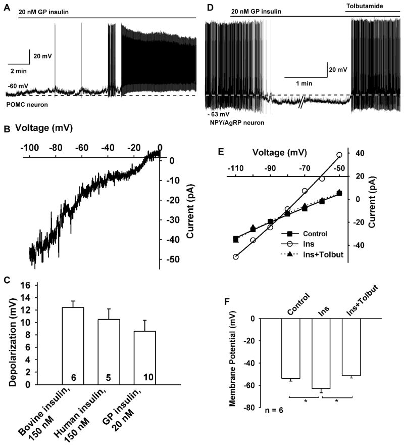
Figure 2. Insulin depolarizes and excites mouse POMC neurons and hyperpolarizes and inhibits NPY/AgRP neurons.
(A) GP insulin (20 nM) depolarized and induced firing in a mouse POMC neuron. (B) The I–V relationship for the insulin-induced current was obtained by digital subtraction of the control I–V from the I–V in the presence of insulin (20 nM) with a Cs+-based internal solution and K+ channel blockers in the extracellular CSF (see the Experimental Procedures). The reversal potential of the nonselective cation current was −10 mV (n=3). The I–V relationship showed a typical doubly rectifying shape. (C) Summary of the effects of bovine insulin (150 nM, Sigma-Aldrich I1882), and human recombinant insulin (150 nM, Sigma-Aldrich I9278) to depolarize mouse POMC neurons in comparison to purified guinea pig insulin (20 nM, Nationl Institutes of Health). (D) GP insulin (20 nM) inhibited the firing and hyperpolarized (−10.7 mV) a NPY/AgRP neuron, and the effects of insulin were antagonized by the KATP channel blocker tolbutamide (200 μM). (E) I/V plot revealed that the insulin-induced outward current had a reversal potential close to EK+. The voltage protocol consisted of 1s steps every 10 mV from −50 to −110 mV (Data not shown). Vhold = −60 mV. (F) Change in the membrane potential of arcuate NPY/AgRP neurons with application of 20 nM guinea pig insulin and after the addition of 200 μM tolbutamide (n=6). Data points represent the mean ± SEM. *p < 0.05.

