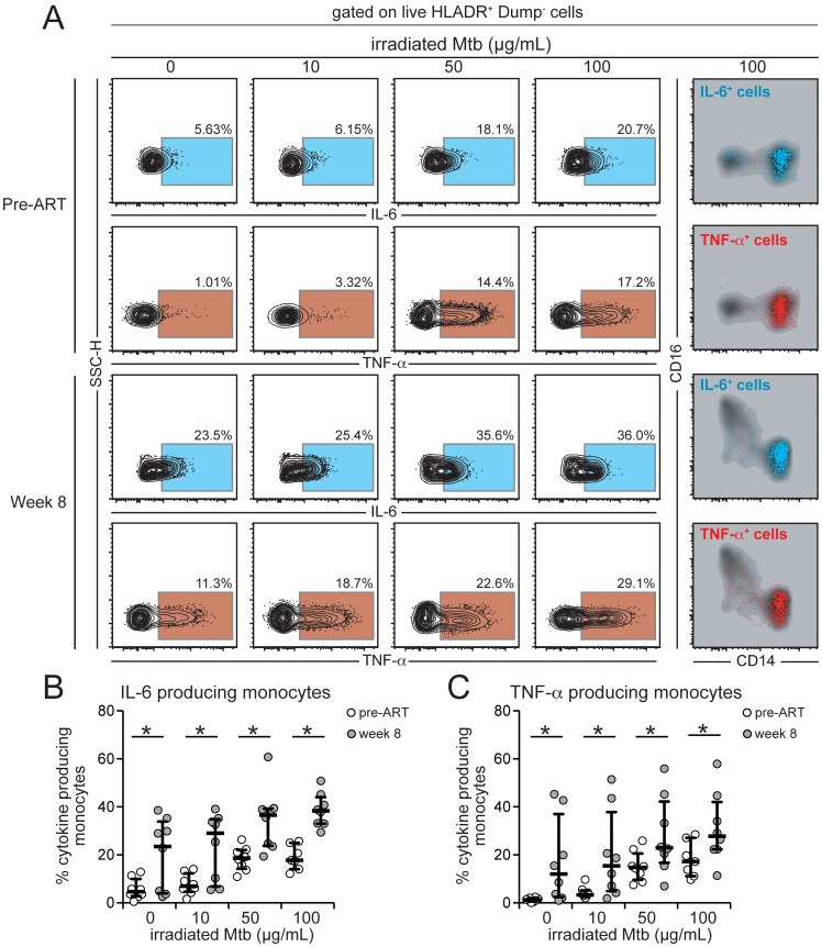
Figure 7. Intracellular production of IL-6 and TNF-α by monocytes from HIV+ patients is affected by HIV plasma viremia and mycobacterial antigen load.
Paired PBMC samples from eight HIV-infected individuals prior to ART initiation and after 8 weeks of treatment when patients achieved virological suppression were incubated in vitro with different doses of irradiated M. tuberculosis (Mtb) for 6 h in the presence of brefeldin-A. Intracellular cytokine assays for detection of IL-6 and TNF-α in monocytes (HLA-DR+Dump− cells) were performed by multicolor flow cytometry. Gates for cytokines were set up based on fluorescent minus one controls. (A) Representative FACS plots of percentage of monocytes expressing IL-6 or TNF-α (left panels) following stimulation with increasing doses of Mtb. Right panel shows overlays of CD14 vs. CD16 expression on cytokine-producing cells after stimulation with irradiated Mtb 100 µg/mL and reveals that most of the cytokine-producing cells in this in vitro system are CD14++CD16− monocytes. (B) Percentage of monocytes from HIV+ patients with high or low viral loads stained positive for intracellular IL-6 after in vitro stimulation with different doses of irradiated Mtb. Data were analyzed using the Wilcoxon matched pairs test. (C) Percentage of monocytes from HIV+ patients with high or low viral loads stained positive for intracellular TNF-α after in vitro stimulation with different doses of irradiated Mtb. Data were analyzed using the Wilcoxon matched pairs test. * P<0.05.

