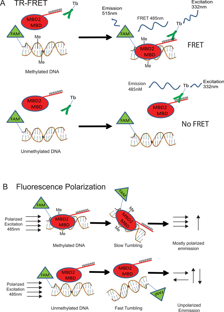
Figure 1. Overview of TR-FRET and Fluorescence Polarization MBD2-MBD DNA-binding assays.
(A) TR-FRET overview: MBD2-MBD protein containing a hexa-histidine tag is mixed with FAM-labeled DNA and terbium-labeled anti-penta-His antibody (Tb-Ab). The MBD2-MBD-Tb-Ab-bound complex is excited with a pulse of 332nm laser light and emission is monitored at 485nm and 515nm (result of FRET) after a 50 µsec delay. The ratio of the 515nm and 485nm emission intensity provides a measure of the extent of binding. (B) Fluorescence polarization assay overview: MBD2-MBD is incubated with FAM-labeled DNA. The reaction is excited with plane-polarized light, and the extent of polarization of the emitted light is measured using parallel and perpendicular polarization emission filters.

