FIGURE 5.
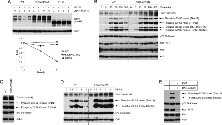
Tiam1 degradation controls the duration of the mitogen-induced mTOR-S6K signaling pathway. A, HEK293 cells expressing HA-tagged wild type Tiam1, HA-tagged Tiam1(S329A/S334A), or HA-tagged Tiam1-C1199 were incubated in low serum for 24 h and then treated with PMA and cycloheximide (CHX) for the indicated times. Cells were collected and lysed. Whole cell lysates were analyzed by immunoblotting with an anti-HA antibody. Actin is shown as a loading control. The graph shows the quantification of Tiam1 abundance relative to the amount at time 0. B, HEK293 cells transduced with retroviruses expressing HA-tagged wild type Tiam1 or HA-tagged Tiam1(S329A/S334A) were incubated in low serum for 48 h and then treated with PMA. At the indicated times, cells were collected and lysed. Whole cell extracts were analyzed by immunoblotting with antibodies for the indicated proteins. Levels of GTP-loaded Rac1 were analyzed in a pulldown assay as described under “Experimental Procedures.” Actin is shown as loading control. To facilitate comparison, a dotted line separates samples from cells expressing wild type Tiam1 and samples from cells expressing Tiam1(S329A/S334A). C, whole cell lysates of asynchronously growing HEK293 cells expressing HA-tagged wild type Tiam1 or HA-tagged Tiam1(S329A/S334A) were analyzed by immunoblotting with antibodies for the indicated proteins. D, as in B except that serum (instead of PMA) was used as mitogen. Cul1 is shown as loading control. E, HEK293 cells were incubated in low serum for 48 h and then treated with PMA. Six hours after PMA treatment, cells were analyzed by immunoblotting. Rac1 activity was assessed as in B.

