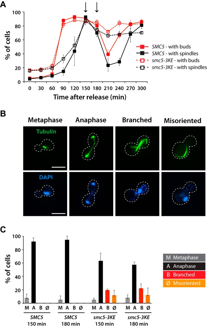
FIGURE 9.

The microtubule-binding mutant of Smc5 exhibits temperature-dependent spindle defects in yeast. A, quantification of cells with buds (red lines) and spindles (black lines) in SMC5 (solid lines) and smc5-3KE (dashed lines) yeast strains after release from G1 arrest at 18 °C. Arrows, time points that were analyzed by immunofluorescence. B, representative mitotic phenotypes observed for smc5-3KE are shown, in comparison with the wild-type morphology (metaphase and anaphase). The peripheries of the yeast cells shown in the pictures are outlined by dashed lines. Scale bars, 5 μm. C, quantification of the different spindle structures observed in mitosis. Data in A and C are shown as averages of three independent experiments; error bars, S.D.
