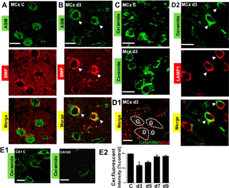
FIGURE 7.
Analysis of ASM activity in the postischemic motor cortex and CA1. The activity of ASM was assessed by colocalization of ASM and BMP, which is an essential co-factor for facilitating ASM activity. A, the absence of colocalization between ASM and BMP in the non-ischemic control motor cortex (MCx C). B, colocalization of ASM and BMP in postischemic day 3 motor cortex (MCx d3). Arrows, colocalization (yellow signal) based on colocalization coefficients. C, confocal microcopy analysis using ceramide antibody (green signal), showing a change in the cellular distribution of ceramide in the postischemic day 3 motor cortex as compared with the non-ischemic control motor cortex. D1, double labeling of ceramide antibody (green signal) with a plasma membrane marker, wheat germ agglutinin (WAG, red signal) in postischemic day 3 motor cortex. The absence of colocalization indicates that ceramide was not mobilized to the plasma membranes. The white interrupted line is used to define the plasma membrane, whereas the solid white line is used to define the nuclear membrane. N, nuclear. D2, colocalization of ceramide (green signal) and lysosomes (LAMP1; red signal) indicates that activation of ASM led to the generation of the lysosomal ceramide in postischemic day 3 motor cortex (MCx d3). Arrows, colocalization (yellow signal) based on colocalization coefficients. E1, confocal microcopy analysis of ceramide levels using ceramide antibody (green signal) in the non-ischemic control CA1 (CA1 C) and the postischemic day 3 CA1 (CA1 d3). E2, quantitative analysis showing that ceramide levels were decreased in the postischemic CA1 at day 3. C, non-ischemic control. d3, d5, d7, and d9, postischemic days 3, 5, 7, and 9. The values represent the mean ± S.E. (error bars) of three independent experiments. Asterisks show significant difference from the non-ischemic control (*, p < 0.05). Scale bar, 20 μm.

