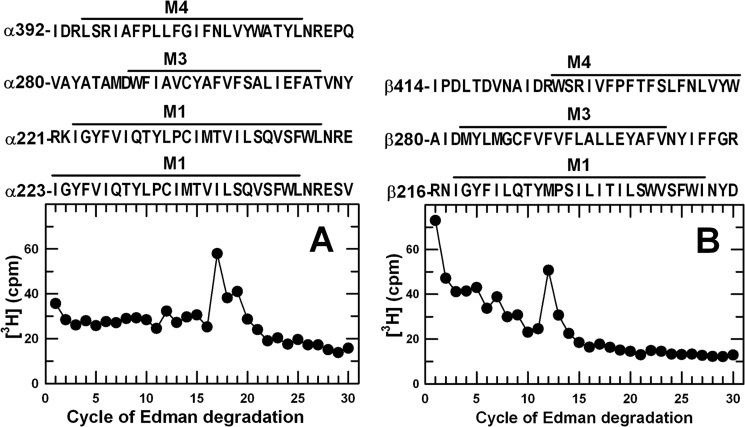
FIGURE 5.
3H release profiles obtained by N-terminal sequence analysis of EndoLys-C digests of α1 and β3 subunits isolated from [3H]AziPm-photolabeled GABAAR. Digests of α1 (A) and β3 (β359 kDa, B) subunits isolated from GABAARs photolabeled with 11 μm [3H]AziPm were loaded directly onto PVDF sequencing filters without prior purification by rpHPLC. Included above each panel are the subunit fragment sequences containing transmembrane helices that can be produced by EndoLys-C digestion. In this experiment, 3,850 cpm of α subunit and 5,125 cpm of β subunit digests (−PPF) were loaded on filters, and 4/5 of the material from each cycle of Edman degradation was collected for determination of released 3H.

