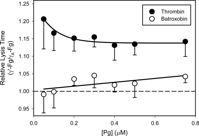
FIGURE 7.
Comparison of lysis times of clots formed from γ′-Fg or γA-Fg with thrombin or batroxobin. γ′-Fg or γA-Fg (7 μm) was incubated at 37 °C with Pg, at concentrations ranging from 0 to 0.75 μm, 5 mm CaCl2, and 400 μm S-2251. Clots were formed by adding the mixtures to wells of a 96-well plate containing either 5 nm thrombin (closed symbols) or 3 units/ml batroxobin (open symbols) and 0.25 nm tPA. Absorbance was monitored at 400 nm, and lysis times were determined as described in the legend for Fig. 2. Each experiment done at least three times, and symbols represent the mean relative lysis times, which were calculated by dividing the lysis times of γ′-Fn clots by those of γA-Fn, while the bars above the symbols reflect S.D. The lines joining the symbols were arbitrary. The dashed line represents normalization ratio of 1, indicating equal lysis times.

