
FIGURE 3.
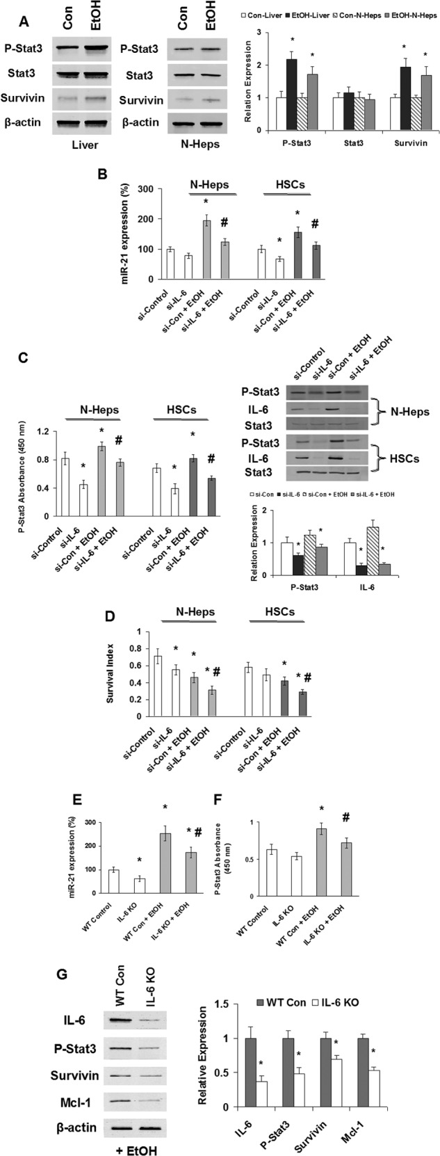
EtOH/IL-6 activation of Stat3 and miR-21. A, Western blots of EtOH mouse liver tissues and N-Hep cell lysates were performed and sequentially probed with antibodies against total-Stat3, p-Stat3, survivin, and β-actin as a loading control in liver tissues or hepatocytes as indicated. Representative immunoblots are shown on the left panel along with quantitative data that show the mean ± S.E. from four separate blots of independent experiments on the right panel. The expression of phospho-Stat3 (Tyr-705) and downstream protein survivin is up-regulated after EtOH treatment in vivo and in vitro. B–D, silencing IL-6 modulates EtOH-dependent miR-21 and Stat3 activation, as well as cell survival in normal human hepatocytes and hepatic stellate cells. B and C, siRNA to IL-6 or control was transfected with or without EtOH treatment (100 mm, 72 h). TaqMan real time assay (B), ELISA (C, left panel), and Western blot analysis (C, right panel) have demonstrated that silencing IL-6 significantly decreased miR-21 expression and Stat3 activation in both cell lines with or without EtOH treatment. C, right panel, representative immunoblots are shown on the top panel along with quantitative data that show the mean ± S.E. from four separate blots of independent experiments on the bottom panel. D, illustrated is the survival index measured by 3-(4,5-dimethylthiazol-2-yl)-5-(3-carboxymethoxyphenyl)-2-(4-sulfonylphenyl)-2H-tetrazolium assay in EtOH-treated N-Heps and HSCs with or without si-IL-6 treatment. Representative and quantitative data (means ± S.E.) from four separate experiments are shown. E and F, IL-6 knock-out mice (IL-6 KO) or wild type control mice (WT Con) were treated with or without EtOH for 5 weeks. TaqMan real time assay (E) and ELISA (F) have demonstrated that silencing IL-6 in vivo significantly decreased miR-21 expression and Stat3 activation by EtOH treatment in mice liver. G, Western blots of EtOH mice liver tissues (with or without IL-6 knock-out) sequentially probed with antibodies against IL-6, p-Stat3 (Tyr-705), survivin, Mcl-1, and β-actin as a loading control as indicated. Representative immunoblots are shown on the left panel along with quantitative data that show the mean ± S.E. from four separate blots of independent experiments on the right panel. The expression of phospho-Stat3 (Tyr-705) and downstream protein survivin/Mcl-1 is down-regulated after IL-6 knock-out in EtOH-treated mice liver in vivo. *, p < 0.05 relative to si-controls or WT controls. #, p < 0.05 relative to si-controls or WT-controls + EtOH.