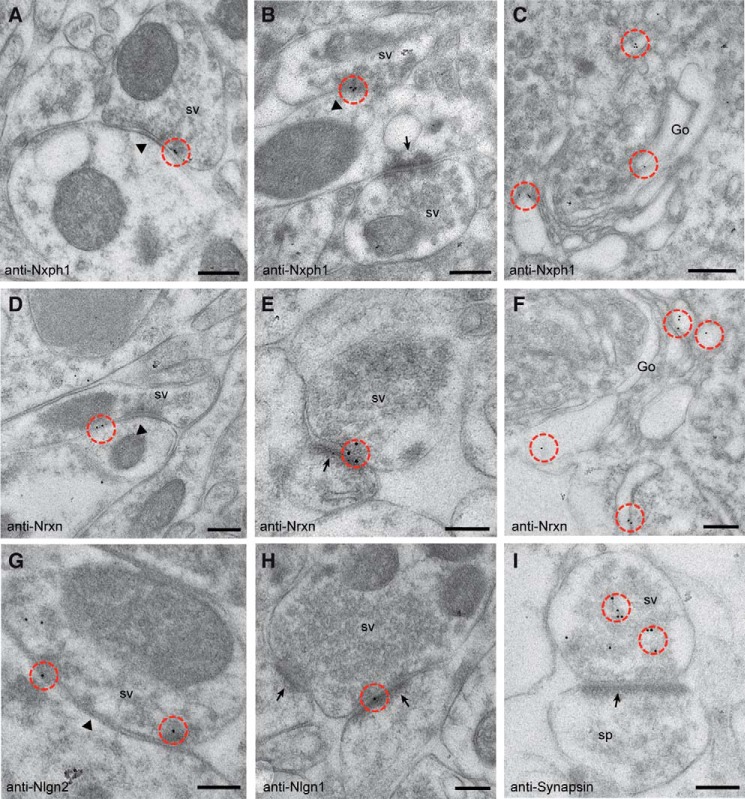
FIGURE 1.
Ultrastructural localization of endogenous Nxph1. Immunoelectron microscopy of Lowicryl-embedded neocortical tissue from murine brain was used to determine the exact localization of Nxph1 (A-C) and its cognate receptor Nrxn (D–F). sv, synaptic vesicle. Post-embedding with 10-nm gold-labeled secondary antibodies reveal Nxph1 only at symmetric, type 2 terminals (A and B, arrowheads), whereas asymmetric, type 1 contacts (B, arrows) are devoid of gold particles (circled in red in all panels). Nrxn is seen at both type 2 (D, arrowhead) and type 1 (E, arrow) synapses, representing inhibitory and excitatory terminals, respectively. C and F, labeling of Nxph1 and Nrxn in Golgi cisternae (Go) demonstrate their passage through the secretory pathway. G–I, control experiments showing the predicted differential distribution of the trans-synaptic Nrxn ligand Nlgn2 at symmetric (G) and Nlgn1 at asymmetric (H) synapses. I, a different labeling pattern is observed with anti-synapsin antibodies, confirming its association with synaptic vesicles in the presynaptic terminal of a type 1 spinous contact (sp). Scale bars, 200 nm, except in C and F, 300 nm.

