
FIGURE 6.
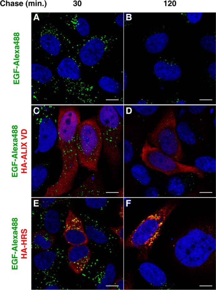
Alix VD expression does not block the EGF degradation pathway. A–F, HeLa cells were grown on coverslips and transfected with control plasmid (empty pCIneo) (A and B), pCIneo 3×-HA-Alix VD (C and D), or pHA-HRS (E and F). The cells were incubated in Opti-MEM media for 1 h followed by incubation with the same media supplemented with Alexa 488-EGF (2 μg/ml) for 30 min at 4 °C. Cells were then incubated at 37 °C for the times indicated above the panels. After incubation, cells were fixed, permeabilized, and stained with mouse monoclonal antibody to HA, followed by Alexa 594-conjugated donkey antibody to mouse IgG (red channel). Nuclei were labeled with DAPI (blue channel). Cells were imaged by confocal laser scanning microscopy. Bars, 10 μm.