FIGURE 2.
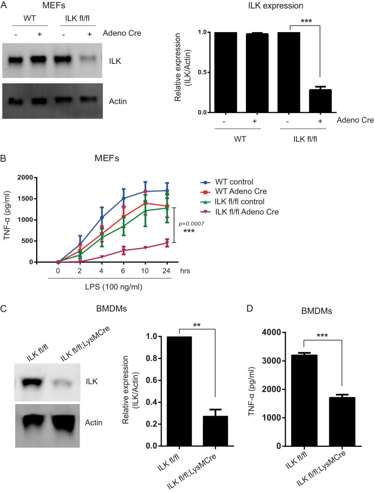
ILK is required for LPS-induced TNF-α production. A, left panel, representative Western blot analysis showing Adeno-Cre-mediated ILK knockdown in ILKfl/fl MEFs. Right panel, densitometric analysis showing the differences in ILK protein bands against actin in MEFs over more than three different experiments. B, time course of LPS-induced TNF-α production in WT and ILKfl/fl MEFs with and without Adeno-Cre virus infection. C, left panel, representative Western blot analysis showing LysM-Cre-mediated ILK deletion in BMDMs. Right panel, densitometric analysis showing the differences in ILK protein bands against actin in BMDMs over more than three different experiments. D, TNF-α production in the supernatants of BMDMs (ILKfl/fl and ILKfl/fl;LysMCre) treated with LPS (100 ng/ml) for 4 h. Data are representative of three different experiments. p values were calculated by two-tailed t test and were relative to LPS stimulation alone: **, 0.001 < p ≤ 0.01; ***, p ≤ 0.001.

