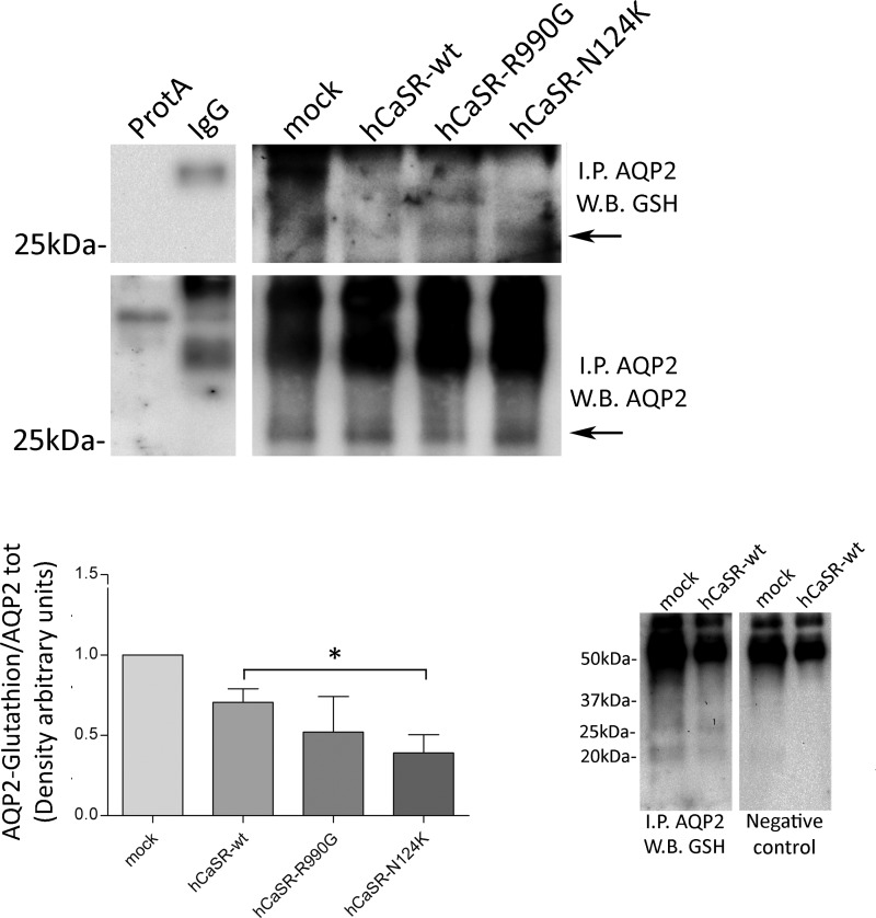
FIGURE 3.
AQP2 was immunoprecipitated (I.P. AQP2) from HEK-293 cells stably expressing hAQP2. Lysates from HEK-293 cells were also subjected to precipitation with only protein A-coupled Sepharose (ProtA) or with nonspecific IgG. Throughout this study, nonequivalents (10 and 90%) of the immunoprecipitates were immunoblotted for AQP2 or GSH, respectively. The data (means ± S.E.) were analyzed by one-way analysis of variance followed by Newman-Keuls multiple comparison test with p < 0.05 (*) considered to be statistically different. AQP2 was immunoprecipitated from mock and hCaSR-wt expressing cells and immunoblotted with or without glutathione antibodies. W.B., Western blot.

