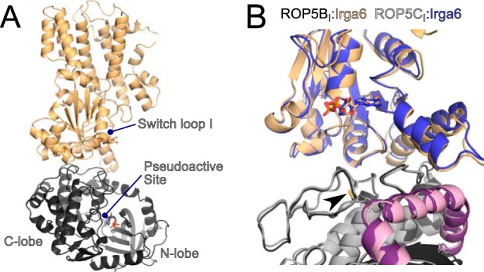
FIGURE 1.

ROP5BI and ROP5CI bind IRGa6 in a similar conformation distal from the pseudokinase and GTPase active sites. A, the structure of IRGa6 (orange) bound to ROP5BI (gray) is shown with their respective bound nucleotides shown in sticks. B, an overlay of the two structures near the ROP5-IRGa6 interface is shown. Cα RMSD for the complex is 1.6 Å over 754 atom pairs, and the Cα RMSD values for the individual IRGa6 and ROP5 chains are 1.1 Å over 398 atom pairs and 0.9 Å over 356 atom pairs, respectively. The ROP5 N-terminal extension is highlighted in pink, and the conserved disulfide bridge (Cys-458/492) that stabilizes the loop in ROP5 that interacts with IRGa6 helix 3 is indicated with an arrowhead.
