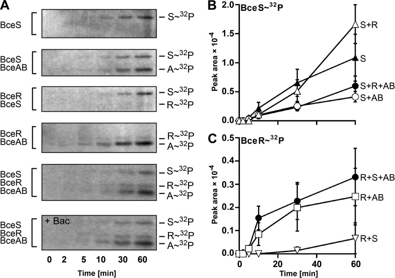
FIGURE 7.
In vitro phosphorylation of Bce module proteins. Purified, detergent-solubilized BceS-His8 was mixed in equimolar ratios with BceAB, BceR, or both as indicated on the left. A, phosphorylation was started at t = 0 min by adding [γ-32P]ATP. At the indicated times, reactions were stopped; the samples were subjected to SDS-PAGE, and phosphorylated proteins were detected by phosphorimaging. Representative autoradiographs of three to four independently performed experiments are shown. B and C, band intensities of BceS-32P (B) and BceR-32P (C) were quantified and plotted over time. The protein combinations in each assay are given on the right by the last letter of their names. Data are shown as the mean ± S.D. of three to four independently performed experiments.

