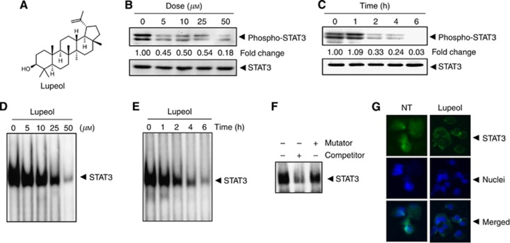
Figure 1.
(A) The chemical structure of lupeol. (B) Lupeol suppresses phospho-STAT3 levels in a dose-dependent manner. HepG2 cells (2 × 10⁶ per ml) were treated with the indicated concentrations of lupeol for 6 h, after which whole-cell extracts were prepared and 30 μg of protein was resolved on 10% SDS–PAGE gel, electrotransferred onto nitrocellulose membranes and probed for phospho-STAT3. The same blots were stripped and reprobed with STAT3 antibody to verify equal protein loading. (C) Lupeol suppresses phospho-STAT3 levels in a time-dependent manner. The HepG2 cells (2 × 10⁶ per ml) were treated with the 50 μM lupeol for the indicated times, after which western blotting was performed as described in (B). The same blots were stripped and reprobed with STAT3 antibody to verify equal protein loading. (D) Lupeol suppresses STAT3 DNA-binding ability in a dose-dependent manner in HepG2 cells. The HepG2 cells were treated with the indicated concentrations of lupeol for 6 h; nuclear extracts were prepared and analysed by EMSA using the radiolabelled oligonucleotide containing hSIE as described in the Materials and Methods. (E) Lupeol suppresses STAT3 DNA-binding ability in HepG2 cells. The HepG2 cells were treated with 50 μM lupeol for indicated time points; nuclear extracts were prepared and analysed by EMSA using the radiolabelled oligonucleotide containing hSIE as described in the Materials and Methods. (F) The EMSA competition assay demonstrating specificity of STAT3 binding. Nuclear extracts of untreated HepG2 cells were prepared and analysed by EMSA using radiolabelled oligonucleotide containing hSIE along with 30-fold excess competitor (an unlabelled oligonucleotide probe) and mutator (an unlabelled probe with a mutation that abrogates STAT3 DNA binding). (G) Lupeol causes inhibition of translocation of STAT3 to the nucleus. The HepG2 cells (1 × 10⁵ per ml) were incubated with or without 50 μM lupeol for 6 h and then analysed for the intracellular distribution of STAT3 by immunocytochemistry. The same slides were counterstained for nuclei with 1 μg ml−1 DAPI for 5 min. The results shown are representative of two independent experiments.

