Figure 2.
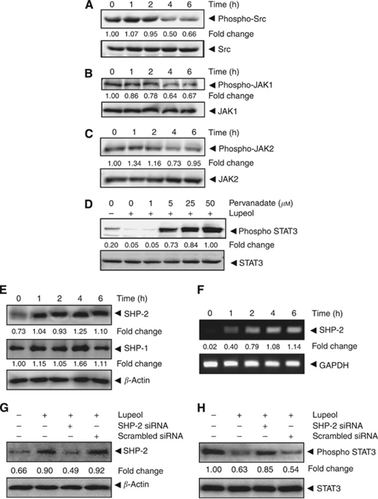
Lupeol inhibits constitutively active STAT3 in HCC cells. (A) Lupeol suppresses phospho-Src levels in a time-dependent manner. The HepG2 cells (2 × 10⁶ per ml) were treated with 50 μM lupeol, after which whole-cell extracts were prepared and 30 μg aliquots of those extracts were resolved on 10% SDS–PAGE, electrotransferred onto nitrocellulose membranes and probed for phospho-src antibody. The same blots were stripped and reprobed with Src antibody to verify equal protein loading. (B) Lupeol suppresses phospho-JAK1 levels in a time-dependent manner. HepG2 cells (2 × 10⁶ per ml) were treated with 50 μM lupeol for indicated time intervals, after which whole-cell extracts were prepared and 30 μg portions of those extracts were resolved on 10% SDS–PAGE, electrotransferred onto nitrocellulose membranes and probed against phospho-JAK1 antibody. The same blots were stripped and reprobed with JAK1 antibody to verify equal protein loading. (C) Lupeol suppresses phospho-JAK2 levels in a time-dependent manner. The HepG2 cells (2 × 10⁶ per ml) were treated with 50 μM lupeol for indicated time intervals, after which whole-cell extracts were prepared and 30 μg portions of those extracts were resolved on 10% SDS–PAGE, electrotransferred onto nitrocellulose membranes and probed against phospho-JAK2 antibody. The same blots were stripped and reprobed with JAK2 antibody to verify equal protein loading. (D) Pervanadate reverses the phospho-STAT3 inhibitory effect of lupeol. The HepG2 cells (2 × 10⁶ per ml) were treated with the indicated concentrations of pervanadate and 50 μM lupeol for 6 h, after which whole-cell extracts were prepared and 30 μg portions of those extracts were resolved on 10% SDS–PAGE gel, electrotransferred onto nitrocellulose membranes and probed for phospho-STAT3 and STAT3. (E) Lupeol induces the expression of SHP-2 protein in HepG2 cells. The HepG2 cells were treated with indicated concentrations of lupeol for 6 h, after which western blotting was performed using SHP-2, SHP-1 and β-actin antibodies. (F) Lupeol induces the expression of SHP-2 in HepG2 cells. The HepG2 cells were treated with indicated concentrations of lupeol for 6 h, after which total RNA was extracted, and the mRNA level of SHP-2 was analysed by RT–PCR. Glyceraldehyde 3-phosphate dehydrogenase (GAPDH) was used as an internal control. (G) Effect of SHP-2 knockdown on lupeol-induced expression of SHP-2. The HepG2 cells were transfected with either SHP-2 siRNA or scrambled siRNA (50 nM). After 24 h, cells were treated with 50 μM lupeol for 6 h and whole-cell extracts were subjected to western blot analysis. (H) The HepG2 cells were transfected with either SHP-2 siRNA or scrambled siRNA (50 nM). After 24 h, cells were treated with 50 μM lupeol for 6 h and whole-cell extracts were subjected to western blot analysis for phosphorylated STAT3. The results shown are representative of two independent experiments.

