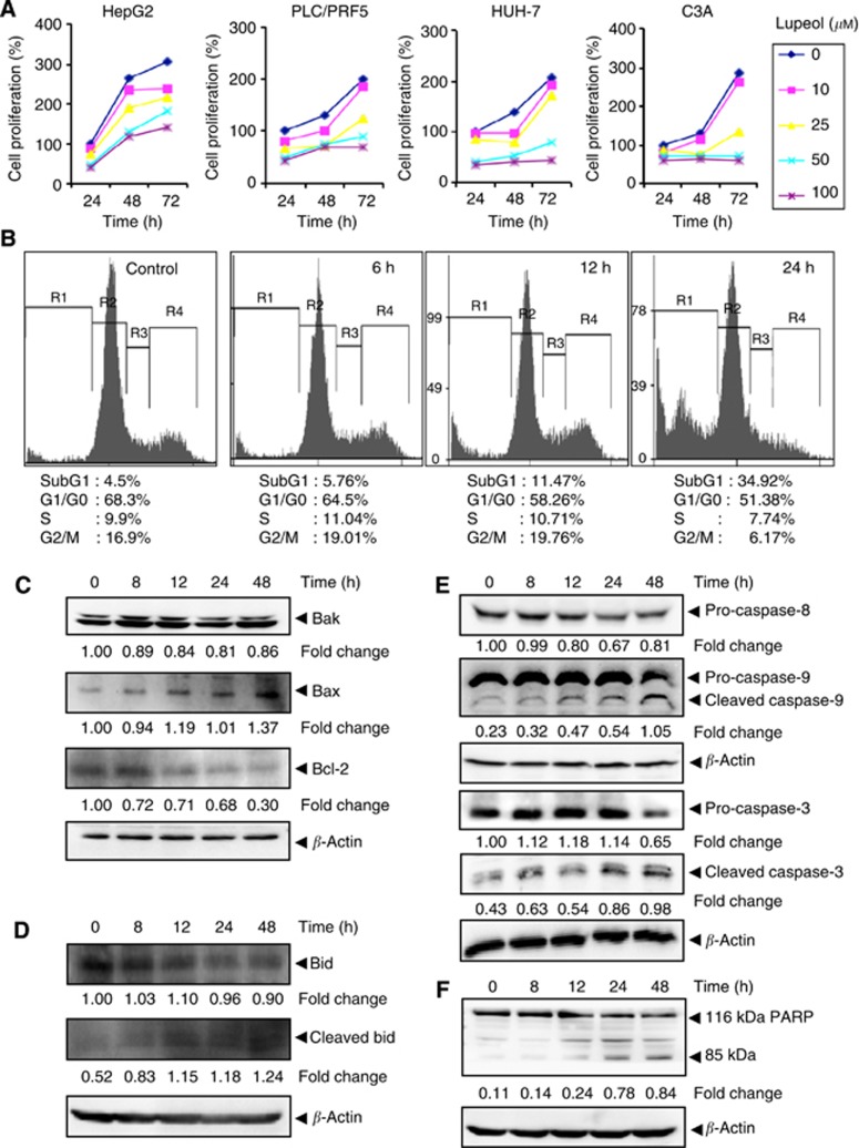
Figure 5.
(A) The HepG2, PLC/PRF5, HUH-7 and C3A cells (5 × 10³ per ml) were plated in triplicate, treated with indicated concentrations of lupeol and then subjected to MTT assay after 24, 48 and 72 h to analyse proliferation of cells. The s.d. values between triplicates are indicated. (B) The HepG2 cells (2 × 10⁶ per ml) were treated with 50 μM lupeol for the indicated times, after which the cells were washed, fixed, stained with PI and analysed for DNA content by flow cytometry. (C) The HepG2 cells were treated with 50 μM lupeol for the indicated times, whole-cell extracts were prepared, separated on SDS–PAGE and subjected to western blotting against Bak, Bax and Bcl-2 antibodies. (D) The HepG2 cells were treated with 50 μM lupeol for the indicated times, whole-cell extracts were prepared, separated on SDS–PAGE and subjected to western blotting against Bid and cleaved Bid antibodies. The same blots were stripped and reprobed with β-actin antibody to show equal protein loading. (E) The HepG2 cells were treated with 50 μM lupeol for the indicated times, whole-cell extracts were prepared, separated on SDS–PAGE and subjected to western blotting against pro-caspase-8, pro-caspase-9, pro-caspase-3 and cleaved caspase-3 antibodies. The same blot was stripped and reprobed with β-actin antibody to show equal protein loading. (F) The HepG2 cells were treated with 50 μM lupeol for the indicated times, and whole-cell extracts were prepared, separated on SDS–PAGE and subjected to western blot against PARP antibody. The same blot was stripped and reprobed with β-actin antibody to show equal protein loading. The results shown are representative of two independent experiments.

