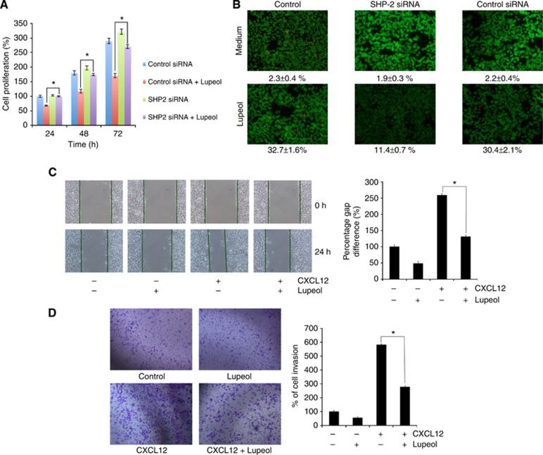
Figure 6.
(A) Gene silencing of SHP-2 abolishes lupeol-induced suppression in tumour cell proliferation. The HepG2 cells were transfected with SHP-2 siRNA or control siRNA for 48 h and then treated with 50 μM lupeol for 24, 48 and 72 h. Mitochondrial dehydrogenase activity was measured at 24, 48 and 72 h after lupeol treatment. Cell proliferation was analysed by measuring absorbance at 570 nm. *P<0.05. (B) Silencing of SHP-2 suppresses lupeol-induced apoptosis. The HepG2 cells were transfected with SHP-2 siRNA or control siRNA for 48 h and treated with 50 μM for 24 h. Apoptosis was measured by live/dead assay. Values represent the mean number of apoptotic cells±s.d. Magnification × 100. (C) Wound-healing assay for evaluating effect of lupeol on the migration of HepG2 cells. An IBIDI culture insert (IBIDI GmbH) consists of two reservoirs separated by a 500 μm thick wall created by a culture insert in a 35 mm Petri dish. For migration assay, an equal number of HepG2 cell (70 μl; 5 × 10⁵ cells per ml) were added into the two reservoirs of the same insert and incubated at 37 °C/5% CO2. After 12 h, the insert was gently removed creating a gap of ∼500 μm. The cells were treated with 50 μM lupeol for 12 h before being exposed to 100 ng ml−1 CXCL12 for 24 h. Width of wound was measured at time 0 and 24 h of incubation with and without lupeol in the absence or presence of CXCL12. The representative photographs showed the same area at time 0 and after 24 h of incubation. (D) Lupeol suppresses invasion of HepG2 cells. The HepG2 cells (2 × 10⁵ cells) were seeded in the top chamber of the Matrigel. After preincubation with or without 50 μM lupeol for 12 h, transwell chambers were then placed into the wells of a 24-well plate, in which we had added either the basal medium only or basal medium containing 100 ng ml−1 CXCL12 in a predetermined arrangement. After incubation for 24 h, cell invasion was analysed and columns represent mean number of invaded cells. *P<0.05.

