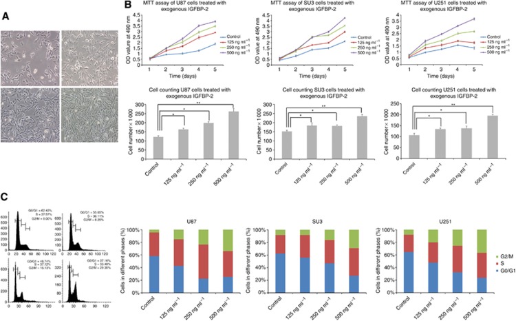
Figure 2.
The effects of exogenous IGFBP-2 on glioblastoma cell proliferation and cell cycle kinetics. (A) Representative photomicrographs of untreated or IGFBP-2-treated U87 cells. Cell proliferation was stimulated by treatment with exogenous IGFBP-2 in a dose-dependent manner. (Upper) left: control; right: 125 ng ml−1 IGFBP-2. (Lower) left: 250 ng ml−1 IGFBP-2; right: 500 ng ml−1 IGFBP-2. (B) Viability of U87, SU3, and U251 cells treated with various concentrations of IGFBP-2, assessed by the MTT assay (upper). Cell quantification following treatment of glioblastoma cells with various concentrations of IGFBP-2 for 48 h (lower). (C) Cell cycle phase was determined from the incorporation of propidium iodide (PI). Cells were seeded in triplicate and analysed 24 h after plating by flow cytometry. The fraction of cells in each phase of the cell cycle is indicated in the graphs. Treatment with IGFBP-2 induced the S- and G2/M-phase entry in a dose-dependent manner. (Upper) left: control; right: 125 ng ml−1 IGFBP-2. (Lower) left: 250 ng ml−1 IGFBP-2; right: 500 ng ml−1 IGFBP-2. Results are presented as mean±s.e. of triplicate samples from three independent experiments. *P<0.05 and **P<0.01.

