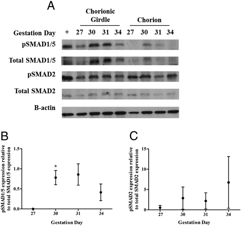
Figure 4.
SMAD signaling protein expression in chorionic girdle and chorion tissue isolated from day 27, 30, 31, and 34 conceptuses. Western blots were probed with antibodies raised against human peptide sequences in SMAD1/5 and pSMAD1/5, or SMAD2, pSMAD2 and murine β-actin (as controls) (panel A). Mouse spleen was used as a positive control tissue (+). Each Western blot was quantified by densitometry (panels B and C). Data were analyzed by repeated-measures ANOVA followed by Tukey multiple-comparison test (*, P ≤ .05) relative to day 27 chorionic girdle (panels B and C). This representative panel shows the pattern of immunoreactive proteins recognized in equine chorionic girdle tissue protein preparations, confirming that the antibodies directed against the human SMAD1/5 and pSMAD 1/5, SMAD2, and pSMAD2 recognized a single equine protein of approximately 60 kDa. Each panel shows typical Western blots in which the tissue-specific protein expression profile was representative of 3 conceptuses.

