
Figure 1.
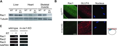
Expression of Rac1 and GLUT4 in skeletal muscle. A) Expression level of Rac1 in liver, heart, and skeletal muscle (gastrocnemius muscle) of wild-type (WT), rac1flox/flox (f/f), and m-rac1-KO (m-KO) mice (8–10 wk old) was analyzed by immunoblotting with an anti-Rac1 antibody. Expression level of tubulin was also analyzed as a loading control. Ten micrograms of protein was loaded on each lane. B) Expression level and subcellular localization of Rac1 and GLUT4 in gastrocnemius muscle fibers of rac1flox/flox and m-rac1-KO mice (8–10 wk old) were analyzed by immunofluorescent staining with specific antibodies. Nuclei were stained with 4′,6-diamidino-2-phenylindole. Images were acquired from the focal plane, as depicted in schematic diagram at right. Blue ellipse in the schematic diagram indicates a nucleus. Scale bar = 20 μm. C) Expression of 3 Rac isoforms (Rac1, Rac2, and Rac3) in gastrocnemius muscle of wild-type and m-rac1-KO mice (8–10 wk old) was analyzed by RT-PCR.