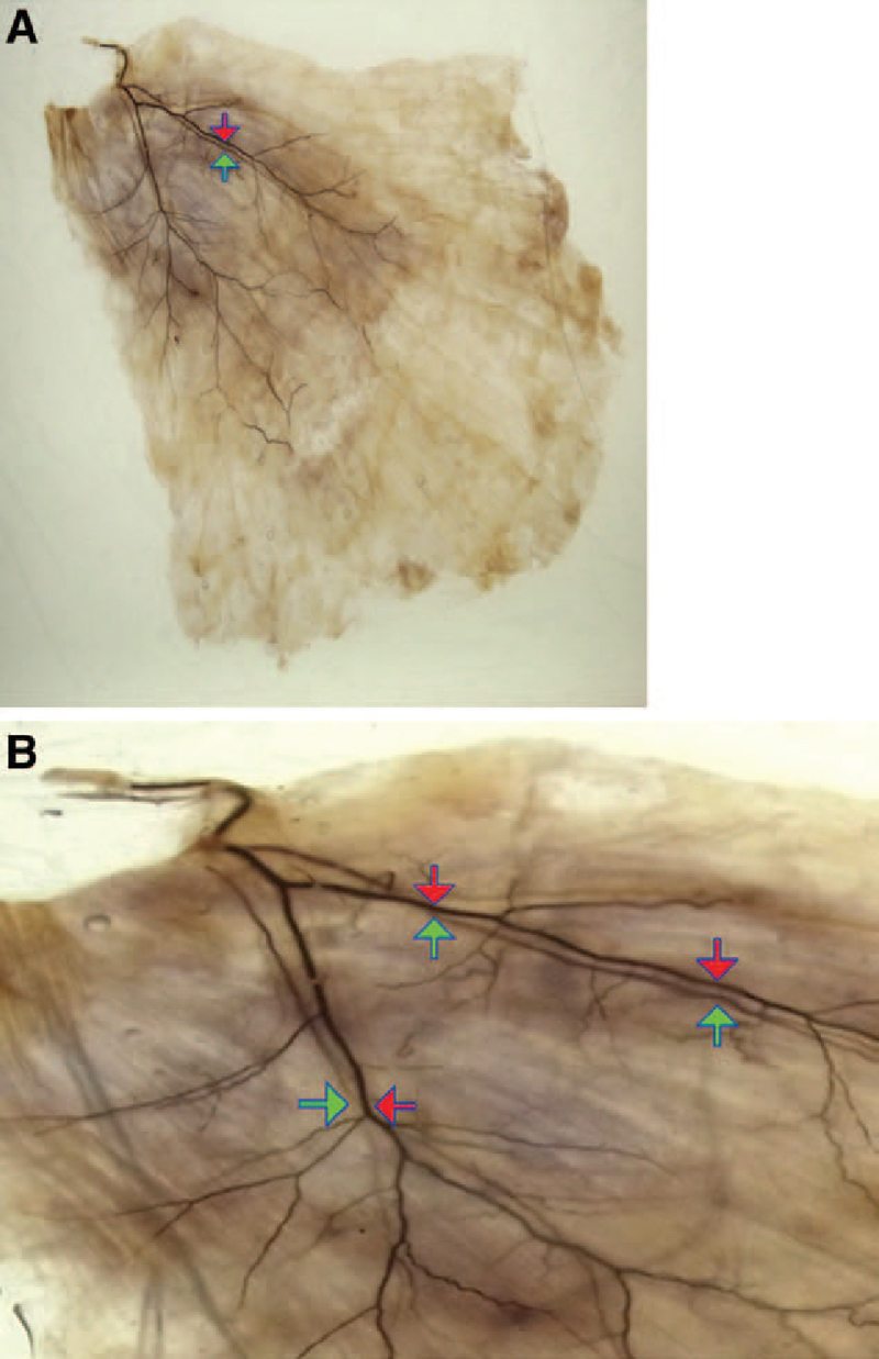
Fig. 3.

The route of the thoracodorsal artery and nerve from a diagonally upward view: The artery and nerve run substantially in parallel to the region in which the periphery may be confirmed. The nerve passes a more superficial layer from the artery after invasion into the muscle. (The red arrow indicates the thoracodorsal artery, and the green arrow indicates the thoracodorsal nerve.) A, The diagonally upward view from the back surface. B, The enlarged image.
