Figure 2.
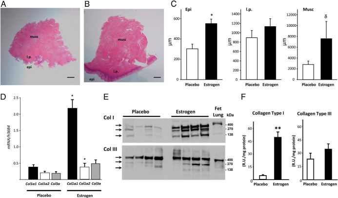
Effect of vaginal estrogen on synthesis activity in the vaginal wall. Hematoxylin and eosin stains are shown; a representative cross-section of vaginal wall from postmenopausal women with prolapse treated with placebo (A) or vaginal estrogen (B) is also shown. Epi, epithelium; l.p., lamina propria; Musc, muscularis. Bar, 1 mm. C, Thickness (micrometers) of epithelium, lamina propria, and muscularis of vaginal tissues from women treated with placebo or estrogen. Data represent mean ± SEM of 12 placebo and eight estrogen. *, P = .002; δ, P = .088. D, Levels of Col1α1, Col 1α2, and Col 3α mRNA in tissues from women treated with placebo (n = 12) or estrogen (n = 8). Data represent mean ± SEM. *, P < .05 compared with the same gene in the placebo group. Immunoblot analysis (E) and densitometry (F) of collagen types I and III (n = 4 per group). Monomeric (140 kDa), dimeric (270 kDa), and trimeric (∼400 kDa) forms of collagen I or collagen III are indicated by arrows. The human fetal lung represents positive control. **, P = .012.

