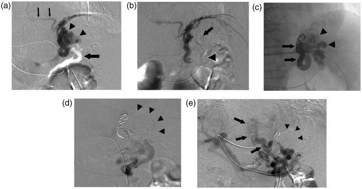
Fig. 1.
A 76-year-old man with a gastric varix locating at the gastric cardia and fornix. (a) Posterior gastric venography revealed that the gastric varix (arrowheads) was fed from one posterior gastric vein (large arrow) and drained into the left inferior phrenic vein (small arrows). (b) The microcatheter was advanced into the drainage vein (arrow) through the gastric varix via the balloon catheter (arrowhead) in the posterior gastric vein. (c) Roentgenogram obtained after embolization of the drainage vein with coils and NBCA (arrows) and injection of 23 mL of 5% EOI showed complete filling of sclerotic agents in the gastric varix (arrowheads). (d) Posterior gastic venography after PTS revealed disappearance of the gastric varix (arrowheads). (e) Splenic venography after PTS revealed that the gastric varix disappeared (arrowheads) and only the flow into the paraesophageal vein without connection with the gastric varix, which was fed from the other posterior gastric vein and the left gastric vein and drained into the azygos venous system (arrows), was evident.

