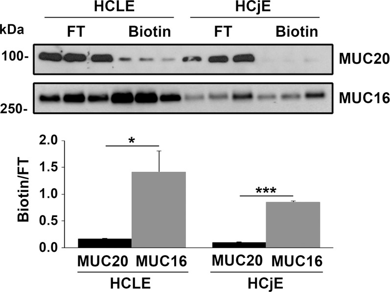
Figure 4.
Protein biotinylation demonstrates low levels of MUC20 at the cell surface. Surface-biotinylated proteins were isolated using a neutravidin-agarose affinity column. By Western blot, MUC20 was detected weakly on the apical surface of stratified HCLE and HCjE cells (upper). In contrast, MUC16 was detected robustly on the apical surface, particularly in HCLE cells. Densitometry analysis of the amount of biotinylated protein normalized to the flow through (FT) demonstrates a higher abundance of MUC16 on the cell surface than MUC20 (lower). Experiments were performed independently in triplicate. *P < 0.05, ***P < 0.001.

