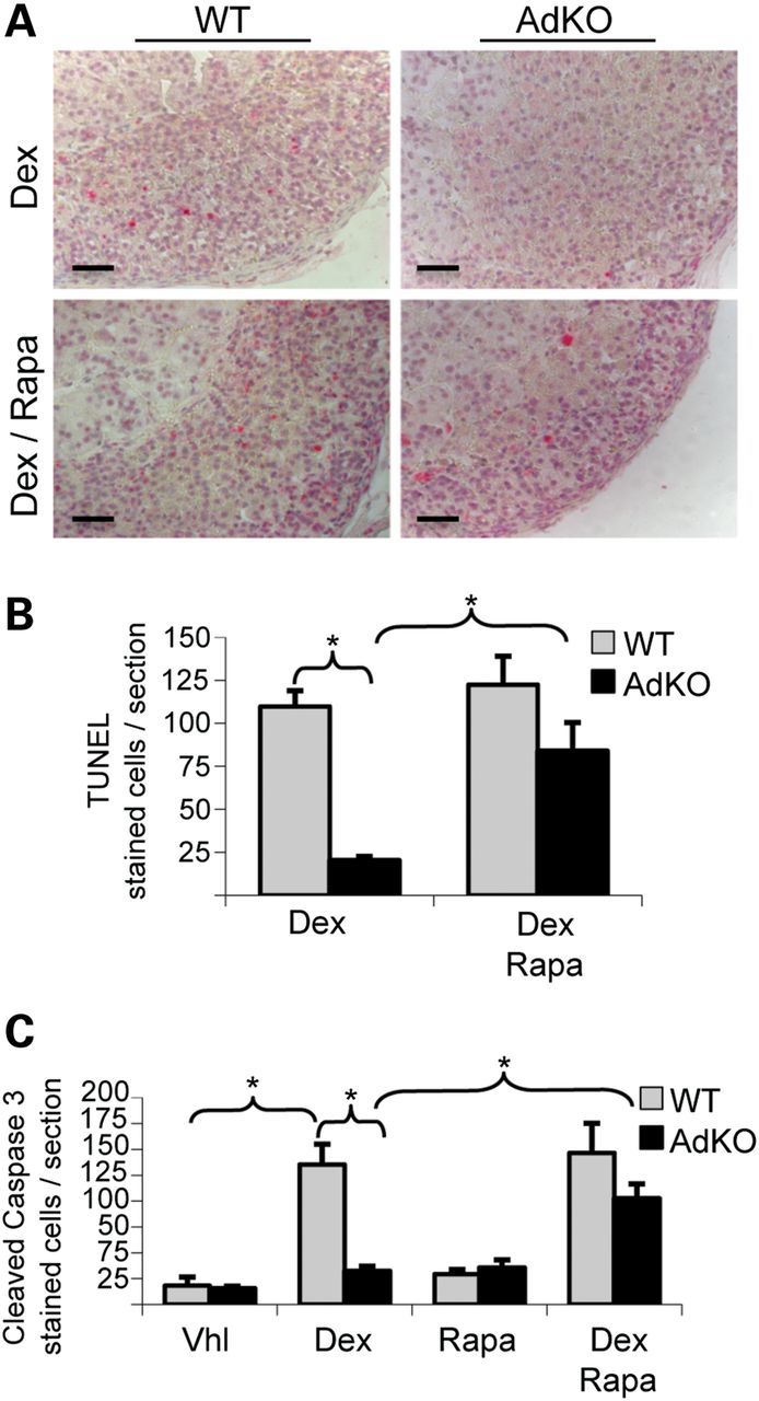
Figure 2.

Dexamethasone-induced apoptosis in adrenals of vehicle or rapamycin-treated WT and AdKO mice. (A) TUNEL staining of adrenal sections from dexamethasone/vehicle-treated WT and AdKO (Dex) compared with dexamethasone/rapamycin-treated WT and AdKO mice (Dex/Rapa). (B) The number of TUNEL-stained cells illustrated in A was quantified in four to seven individuals per condition (values ± SEM). Values represent the number of stained cells per adrenal section expressed as a percentage of the mean of WT. *P < 0.05. (C) Cleaved-caspase 3 staining in adrenal sections was also performed in four different treatment groups (vehicle, dexamethasone, rapamycin, dexamethasone/rapamycin) in both genotypes (WT and AdKO): the number of stained cells was quantified in four to eight individuals per condition (values ± SEM). Values represent the number of cleaved-caspase 3 stained cells per adrenal section expressed as a percentage of the mean of WT. *P < 0.05. Vhl, vehicle; Dex, dexamethasone; Rapa, Rapamycin. Scale bars, 100 µm.
