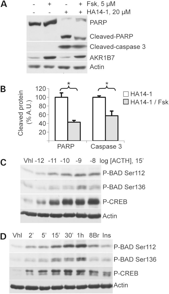
Figure 5.

Apoptosis induction and BAD phosphorylation in murine ATC7 adrenocortical cells. (A) PARP and Caspase 3 cleavage and AKR1B7 levels were detected in ATC7 cells treated with PKA pathway activator forskolin (Fsk) and/or apoptosis inductor HA14-1 for 4 h. (B) PARP and Caspase 3 cleavage was quantified in response to HA14-1 in the presence or absence of forskolin (values ± SEM). Values represent relative band density over actin for cleaved-caspase 3 and relative cleaved protein over total protein for cleaved-PARP, expressed as a percentage of the mean of the control. n = 5; *P < 0.05. Vhl, vehicle. (C) BAD phosphorylation at Ser112 or Ser136 positions was detected in cells treated for 15′ with increasing concentrations of ACTH. (D) BAD phosphorylation at Ser112 or Ser136 positions was detected in cells treated for various amounts of time with the PKA pathway activator ACTH or for 15′ with either the specific PKA activator 8-Br-cAMP (8Br) or PI3K/AKT/mTOR pathway activator insulin (Ins). Total BAD and CREB protein signals were unaffected by these short-time treatments (not shown).
