Figure 1.
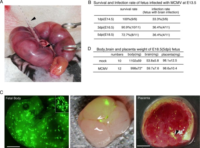
Intraplacental MCMV infection and development of MCMV-infected fetuses. (A) Inoculation procedure during surgery. The arrowhead and arrow indicate the glass micropipette for injection and the labyrinth region in the placenta, respectively. (B) Survival rates of fetuses infected with MCMV at E13.5 and infection rates of their brains. (C) Fluorescence stereomicroscopic views of MCMV-EGFP infectious foci at 3 dpi in a fetus, its brain, and placenta. A magnified view of an infectious focus in the fetus is also shown in the inner panel. The fluorescence images of brain and placenta were superimposed on their bright field images. The arrowhead in the image of the placenta indicates the injection site. Fetal body, scale bar = 2 mm. Brain and placenta, scale bar = 1 mm. (D) Weights of the fetal body, brain, and placenta at E18.5 (5 dpi). Mean ± standard error of mean (SEM) are shown. *P < 0.05 compared with mock-infected mice. MCMV, murine cytomegalovirus; EGFP, enhanced green fluorescent protein.

