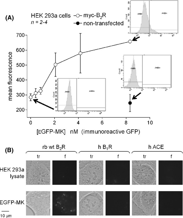
Figure 11.

(A) Cytofluorometry of HEK 293a cells that optionally expressed myc-B2R, sequentially detached and stained for 30 min (37°C) with EGFP-MK recovered from the cell lysate of producer cells (concentration indicated as nmol/L of immunoreactive GFP). (B) Selectivity of HEK 293a cell labeling by EGFP-MK (4.2 nmol/L, 30 min, 37°C; control HEK 293a lysate contained 0.15 nmol/L EGFP) as a function of the expressed transgene (rabbit wild-type B2R, human kinin B1 receptor, human angiotensin-converting enzyme). Matched transmission (tr) and green fluorescence (f) fields are shown side by side (1000×).
