Figure 2.

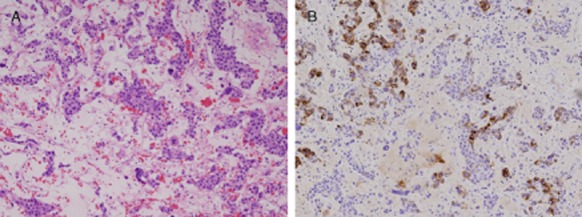
Photomicrograph of needle biopsy specimen. Hematoxylin-eosin stained sections revealed diffuse malignant cells (A 100×) and immunostaining was positive for anti–alpha-fetoprotein (B 100×).

Photomicrograph of needle biopsy specimen. Hematoxylin-eosin stained sections revealed diffuse malignant cells (A 100×) and immunostaining was positive for anti–alpha-fetoprotein (B 100×).