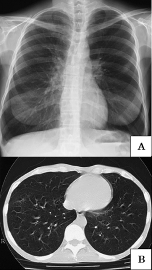
Figure 1.

(A) Chest X-ray on admission. Slight hyperinflation and hyperlucency of both lung fields were observed. (B) Chest computed tomography scan in the inspiratory phase. It shows hyperinflation of both lungs and diminished vascular shadows on peripheral lung fields. Other typical signs of bronchiolitis obliterans, that is mosaic perfusion and air trapping sign, were not observed.
