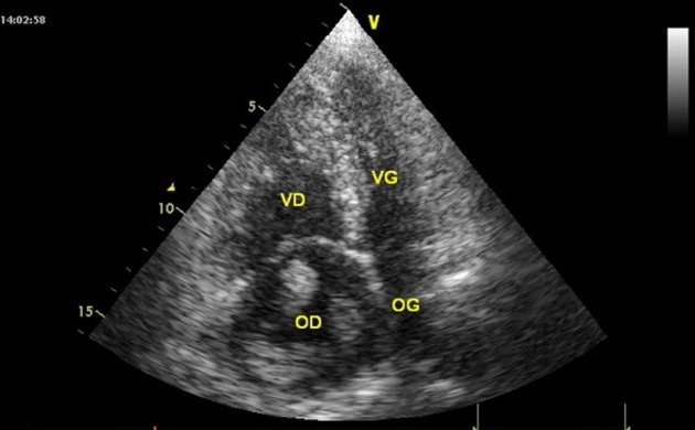
Figure 1.

A transthoracic echocardiographic image; apical four-chamber view showing a dilatation of right chambers and a voluminous heterogeneous, polylobulated horseshoe-shaped mass, in the right atrium.

A transthoracic echocardiographic image; apical four-chamber view showing a dilatation of right chambers and a voluminous heterogeneous, polylobulated horseshoe-shaped mass, in the right atrium.