
Figure 1.
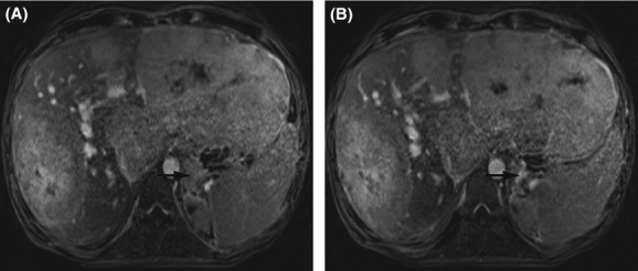
Magnetic resonance imaging of the liver showing hepatomegaly and multiple tumors in the left and right liver lobe before (A) and after 7 months of sorafenib (B). Axial T1-weighted delayed contrast-enhanced images by volumetric interpolated breath-hold examination are shown in portal venous phase of acquisition. Note the left adrenal metastasis (arrows).