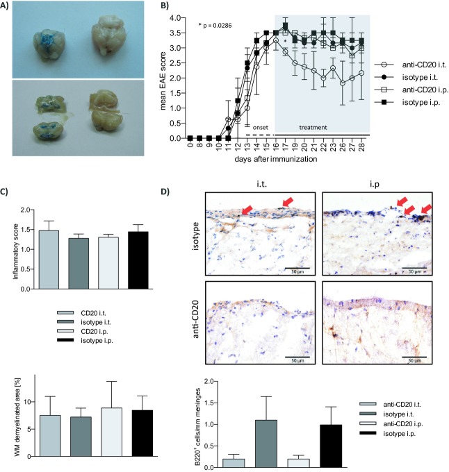
Figure 1.
Intrathecal anti-CD20 effectively depletes meningeal B cells and moderately ameliorates chronic EAE. (A) 10 μL methylene blue dye suboccipitally injected into the cisterna magna accurately distributes in basal cisterns (upper left panel) and ventricles (lower left panel, 1 h post injection; right panels: identical anatomical regions in an untreated control). (B–D) Six- to eight-week-old female C57BL/6 mice were immunized with recombinant MOG 1-117. After individual EAE onset (EAE score ≥2) mice were randomized to receive weekly intrathecal (i.t.) or intraperitoneal (i.p.) injections with 100 μg of murine anti-CD20 or isotype control antibody. (B) Clinical EAE score of n = 12 mice. *P = 0.0286 (anti-CD20 i.t. vs. isotype i.t.; Mann–Whitney test), error bars represent SEM. (C) One week after the last of three weekly injections, the CNS of n = 12 mice were dissected for histologic assessment. For analysis of CNS inflammation and demyelination, brains and spinal cords were sectioned following cryofixation and stained with Luxol fast blue and Periodic acid Schiff (LFB/PAS) or hematoxylin and eosin (H&E). Inflammatory infiltration (H&E; top) was scored as follows: 0 = no inflammation, 1 = little inflammation, 2 = moderate inflammation, 3 = strong inflammation. Demyelination (LFB/PAS; bottom) was evaluated as % of demyelinated area of the entire white matter. Error bars represent SEM. (D) For quantification of meningeal B cells, brain and spinal cord sections were stained by B220 immunohistochemistry. Representative slides are shown. Infiltrating B cells (B220) were counted and graphed as cells per mm meninges. Error bars represent SEM. EAE, experimental autoimmune encephalomyelitis; MOG, myelin oligodendrocyte glycoprotein; CNS, central nervous system.

