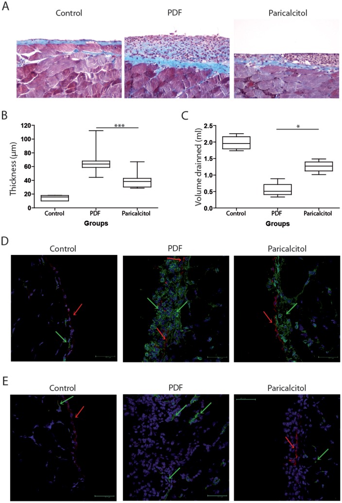
Figure 1. Paricalcitol reduced peritoneal membrane fibrosis, inflammation and ultrafiltration failure in mice exposed to PDF.
A) Paraffin sections of the peritoneal membrane from the 3 groups were stained with Masson's trichrome. B) Thickening of the peritoneal membrane was determined by morphometric analysis. C) Peritoneal permeability was determined by net ultrafiltration. D) The presence of inflammatory and mesothelial cells was determined by the expression of CD45 (green) and cytokeratin (red), respectively, in frozen sections of peritoneal membrane representative of each group. A green arrow indicates hematopoietic cells. A red arrow indicates mesothelial cells. E) The angiogenesis was determined by the expression of CD31 (green). Cytokeratin-positive cells are stained in red. The color balance was equally adjusted in immunofluorescence using Photoshop V10 for Mac (Abobe Systems Incorporated, US). n≥5 in each group. Statistical significance was determined using the Mann-Whitney test. *P<.05; ***P<.001.

