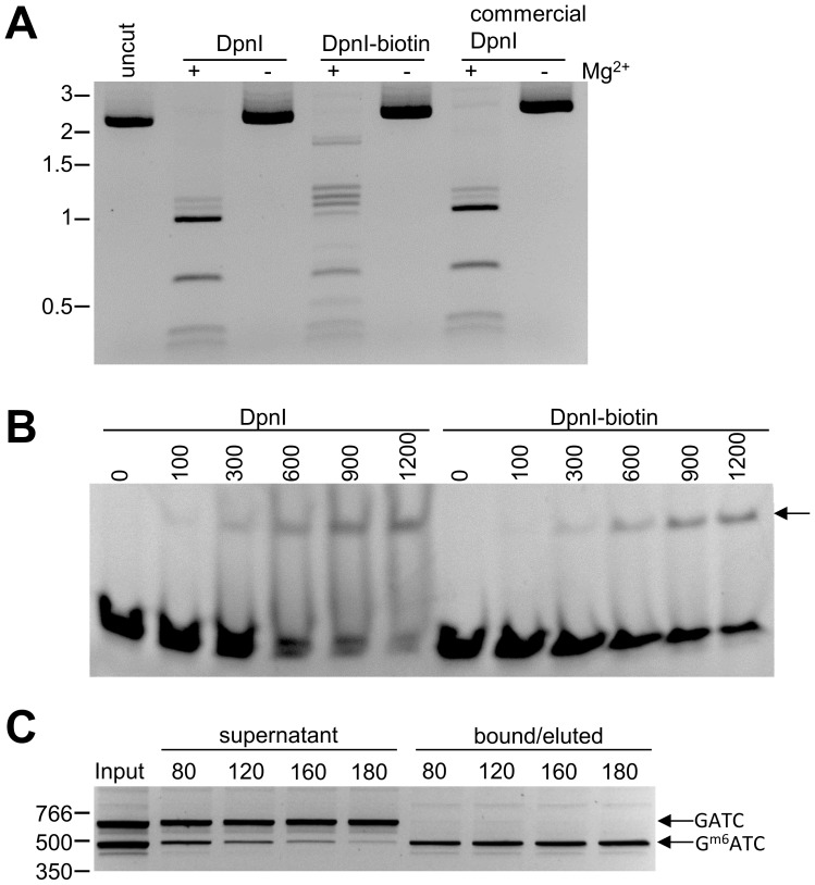
Figure 1. Analysis of biotinylated DpnI.
(A) pUC19 was incubated with DpnI, DpnI-biotin or commercially sourced DpnI in the presence or absence of 10 mM magnesium chloride. The digested fragments were separated on a 1.5% agarose gel. (B) A FAM-labeled DNA duplex containing one Gm6ATC site was incubated with increasing amounts of DpnI or DpnI-biotin (0 to 1200 ng). The reactions were separated on a 20% TBE gel and analyzed with fluorescence imaging. (C) An unmethylated 651 bp DNA fragment and a Dam-methylated 477 bp DNA fragment were combined and incubated with increasing amounts of immobilized DpnI-biotin (80–180 µl). DNA was eluted using GTC and desalted. All fractions were separated on a 3% agarose gel.

