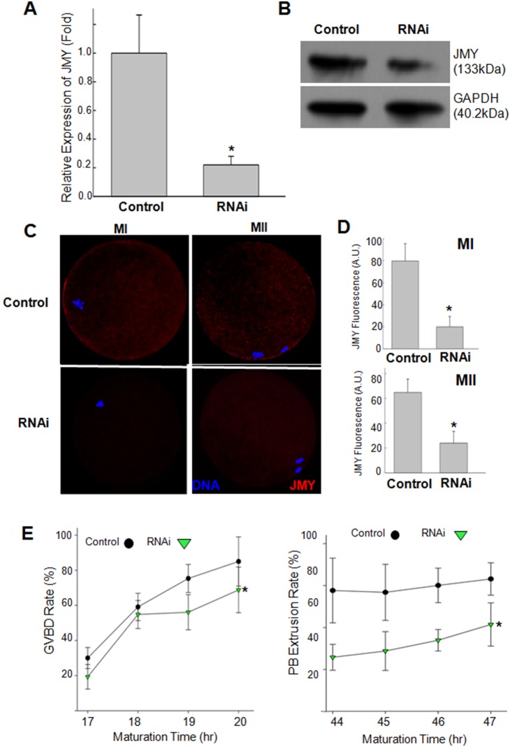
Figure 2. Knockdown of JMY during meiotic maturation.
A–D: GV oocytes injected with dsRNAs or control were cultured for 44 hours. Knockdown of JMY mRNA was determined by RT-PCR (A) and Western blotting (B). Subcellular localization (C) and quantitized fluorescence intensity (D) of JMY fluorescence of dsRNA or control injected oocytes measured at MI (20 h after culture) and MII (44 h after culture) stages respectively. E: Germinal vesicle breakdown (GVBD) and polar body extrusion (PBE) rates of JMY dsRNA induced oocytes. Red: JMY, Blue: chromatin. Values represent mean ± s.e.m. *p<0.05, n = 5.

