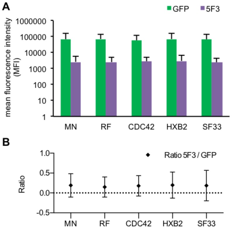
Figure 3. Expression levels of chimeric Env/V3 and GFP.
HEK293T (3×10⁵) cells were separately transduced at MOI 1 by one of the pQL9 derived transduction competent particles encoding one of the chimeric Env/V3-variants, respectively. 72 h after transduction, cells were harvested and stained with an APC-labeled 5F3 antibody binding a constant region within the extracellular gp41 mojety and compared to GFP mediated fluorescence (Pearson correlation p<0,01 for each variant). A FACS analyses are depicted as the mean fluorescence intensity (MFI) of APC labeled 5F3 antibody- and GFP-signals for all chimeric Env/V3 variants, respectively. B The MFI ratios of 5F3 to GFP signals were calculated, respectively.

