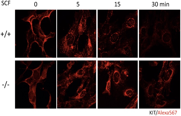
Figure 4. Localization of KIT after SCF stimulation is altered in CALM −/− MEFs.
Localization of KIT was analyzed before and after SCF stimulation under confocal microscopy using WT and CALM −/− MEFs engineered to express KIT. KIT was visualized by the biotinylated anti-KIT antibody (Ab) and AlexaFluor 568 streptavidin conjugates.

