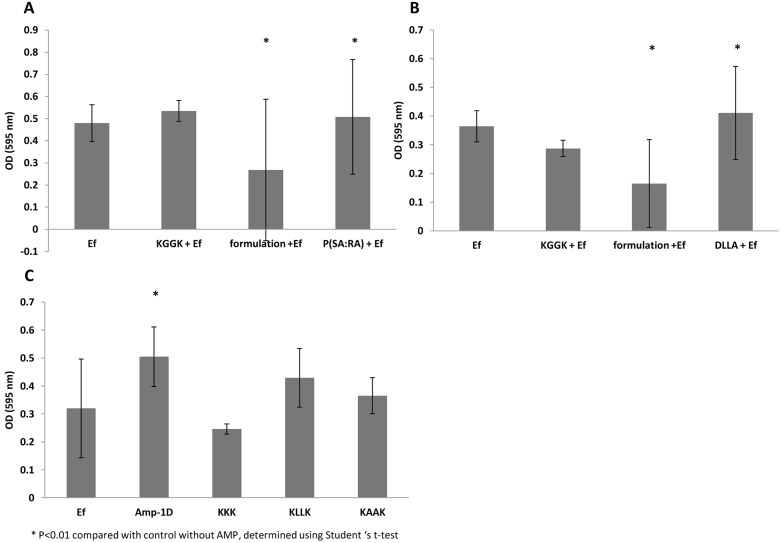
Figure 3. Effect of the antimicrobial peptides on the development of E. faecalis biofilms.
E. faecalis biofilms were grown in 96 microtiter plate wells for 72 hrs in the presence of KGGK formulated with P(SA:RA) (panel A), or formulated with DLLA (B,) or with soluble peptides (C). Ef represents the non-treated bacteria, KGGK+EF - bacteria treated only with peptide, formulation+EF - bacteria treated with polymer and peptide and Ef + polymer - bacteria treated only with polymer control). The biofilm was stained with 1% crystal violet measured at OD 595 nm (see Materials and Methods). The optical density of the polymers alone without the bacteria was subtracted from the results of the biofilm that came in contact with the formulation and the polymer.

