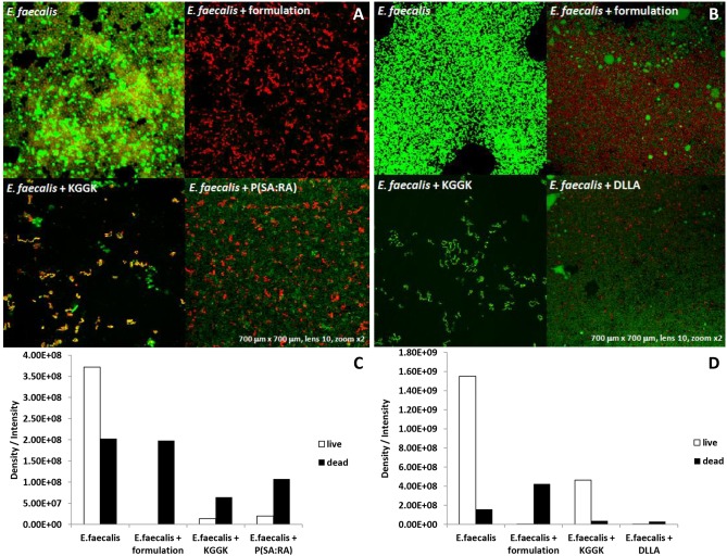
Figure 5. Live/dead assay.
E. faecalis came in contact with the examined materials for 72 hrs to form biofilm. The medium was discarded and the wells were washed gently with PBS. The live bacteria were stained with green dye, the dead bacteria were stained with a red dye. A 5 ml volume of each dye from the dead/live dying kit was added to 450 µl PBS using an Eppendorf and 30 µl of the solution were added in each well. Images were taken using an Olympus confocal microscope [A, B]. The black column represents the dead bacteria, the white column represents the live bacteria. The biofilm was quantified by measuring the area occupied by the bacteria with the aid of Image Pro 4.5 software (Media Cybernetics) [C, D].

