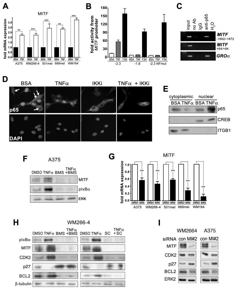
Figure 2. TNFα regulates MITF expression through IKK.
A. Real time qPCR analysis of a panel of melanoma cell lines treated with 50ng TNFα for 24hrs. B. Different MITF promoter construct activity as detected by luciferase in WM266-4 cells treated with 50ng TNFα for 24hrs. Forskolin (FSK) served as positive control. C. NF-κB/p65 Chromatin-IP from TNFα treated WM266-4 cells. The indicated regions of the M-MITF promoter region or a coding region of the MITF gene were amplified. Amplification of the GROα promoter served as positive control (50). D. Immunofluorescence analysis for NF-kB/p65 in WM266-4 cells treated with 50ng TNFα for 2hrs or 0.5μM BMS-345541 for 2hrs either alone or in combination. E. Western blot of cytoplasmic and nuclear extracts from WM266-4 cells treated with BSAE or 50ng TNFα. F. Western blot of A375 cells treated with TNFα or DMSO and 0.5μM BMS-345541 (IKKi) as indicated for 24hrs. G. Real time qPCR analysis of a panel of melanoma cell lines either untreated or treated with 0.5μM BMS-345541 for 24hrs. H. Western blot of WM266-4 melanoma cells treated with TNFα or DMSO, BMS-345541 (0.5μM) or SC-514 (1μM) as indicated for 24hrs. I. Western blot of WM266-4 and A375 melanoma cells transfected with control or MITF specific siRNAs for 24hrs for MITF, CDK2, p27, BCL2 and ERK2.

