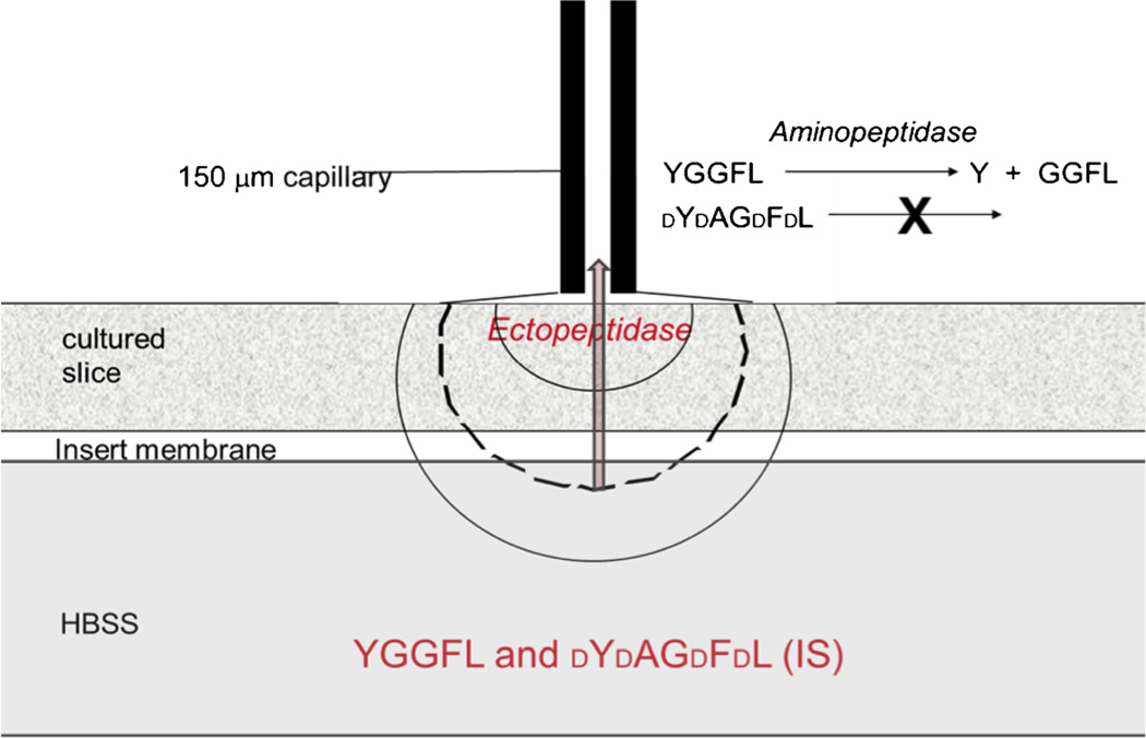
Fig. 4.
Schematic portrayal of the single-probe system. An electrolyte-filled fused-silica capillary is placed perpendicular to the tissue culture and separated from it by a thin, liquid layer (25–50 µm). An electrode and the distal end of the capillary are placed in electrolyte. The other electrode is placed in the HBSS. Peptide substrate and a d-amino-acid peptide are dissolved in the HBSS. The approximately-semicircular lines indicate isopotentials

