
Figure 1.
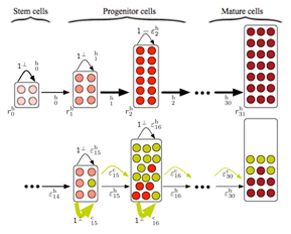
Schematics of our hierarchical hematopoiesis model. At the top, undisturbed healthy hematopoiesis. At the root of our hierarchical structure (compartment 0) is the stem cell compartment with a few slowly dividing stem cells (light red, proliferation rate ) that can self renew with probability and differentiate with probability . Cells undergo several differentiations with probabilities and proliferate with rates until they reach the mature cell state (compartment 31 in our case, dark red). At the bottom, hematopoiesis is disturbed by leukemic cells (dark yellow). The leukemia driving mutation occurs in compartment 15. The self-renewal capacity ( ) of the leukemic cells is significantly increased and malignant cells accumulate in bone marrow compartments. Concomitantly, the proliferation of healthy cells (red) is suppressed by cancer cells and thus the healthy cell count decreases in compartments downstream of compartment 15.