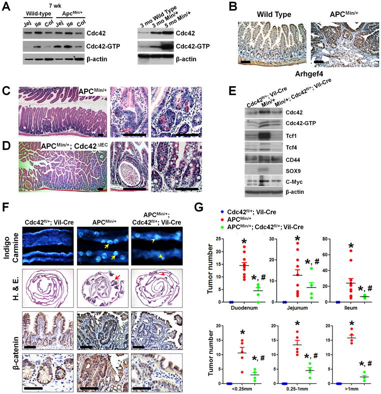
Figure 2. Cdc42 reduction alleviated APCMin/+ intestinal polyposis.
(A) Western blots showed increased levels of active Cdc42 in small and large intestines of 7-week APCMin/+ mouse intestines compared with age matched wild type mice of same genetic background (left panel, compare lanes 1–3 to lanes 4–6). Total and active Cdc42 were increased in older APCMin/+ mice (right panel). Jej: jejunum; Ile: ileum; Col: colon.
(B) Arhgef4 immunohistochemstry showed expansion of its expression domain into adenomatous tissues in APCMin/+ intestines.
(C) Dysplastic lesion in 40-day old APCMin/+ mice. Note that the distorted glands possess large eosinophilic granular debris, typically found in.Paneth cells.
(D) Total deletion of Cdc42 in Cdc42ΔIEC; APCMin/+ mice caused epithelial cystic formations. No dysplastic crypt was found. There is a complete loss of Paneth cell granule in crypts.
(E) Western blots for total, active Cdc42, and several Wnt targets. Note that Cdc42fl/+;Vil-Cre littermate mice, phenotypically wild type in terms of intestinal development and function (19), were used as reference.
(F) Cdc42 reduction decreased the tumor load in APCMin/+ mouse intestines. Macroscopic polyp detection by indigo carmine staining was followed by histology analyses and β-catenin immunohistochemistry on swiss-roll intestinal sections to identify adenoma. Note that Cdc42 inhibition reduced β-catenin nuclear but not epithelial junction staining. All comparison was made from same intestinal segments from littermate animals unless specified otherwise.
(G) Tumor numbers were counted and sizes were measured in duodenum, jejunum and ileum of mice with indicated genotypes. * indicates p<0.05 when compared to Cdc42L/+;Vil-Cre; and # indicates p<0.05 when compared to APCMin/+ mice. Scale bars, 5 μm.

