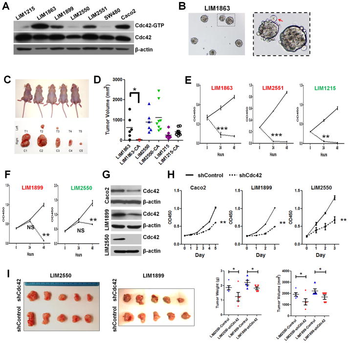
Figure 6. Human CRCs with higher Cdc42 activities were sensitive to Cdc42 inhibition.
(A) Western blots showed different total and active Cdc42 levels in a panel of human CRC cells.
(B) LIM1863 cells grew as suspending organoids in liquid medium.
(C) LIM1863 treated by 10 μM CASIN for 16 hrs showed reduced tumorigenicity in xenograft model. The left and right body flanks of the same nude mice were injected with equivalent numbers of CASIN-treated (T) versus control (C) cells. Mice were imaged at day 70 after cell implantation. Tumors were dissected and photographed from corresponding mice.
(D) Tumor volumes from DMSO and CASIN-treated LIM1863-CA, LIM2550-CA and LIM1215-CA cells were graphed for individual tumors. * indicates p<0.05. Note that LIM2550 and LIM1215 tolerated the transient CASIN treatment.
(E–F) CASIN caused an immediate growth arrest in some CRCs (E) but a delayed inhibition in others (F). Cell lines with relatively high Cdc42 activities were shown in red, otherwise in green.
(G–H) Stable knockdown of Cdc42 by lentiviral shRNA particles caused growth inhibition in vitro.
(I) LIM2550 and LIM1899 cells with stable Cdc42 knockdown showed significantly reduced tumorigenicity in xenograft assays, compared to control knockdown cells (p<0.05 for both tumor volume and tumor weight). Tumors were harvested at day 30 after injection for analyses.

