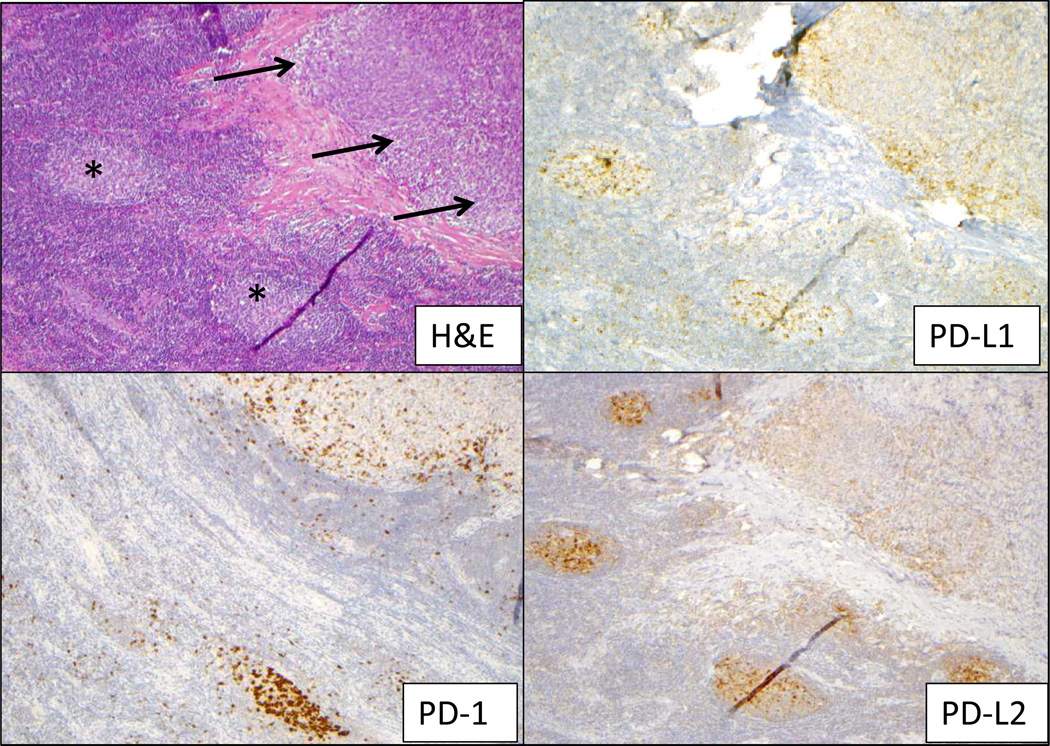
Figure 2. Immunoarchitechture within a melanoma lymph nodal metastasis.
On a section stained with H&E (upper left), a tumor deposit is indicated by arrows and lymph node germinal centers by asterisks. Expression of PD-1, PD-L1 and PD-L2 was observed in the lymph node germinal centers, providing an internal positive control for staining. Within the tumor deposit, PD-L1 and PD-L2 were expressed by both tumor and infiltrating immune cells, associated geographically with PD-1 expression. Additional characterization of the immune infiltrate is provided in Supplementary Figure 4. Original magnification 100×, all panels.

