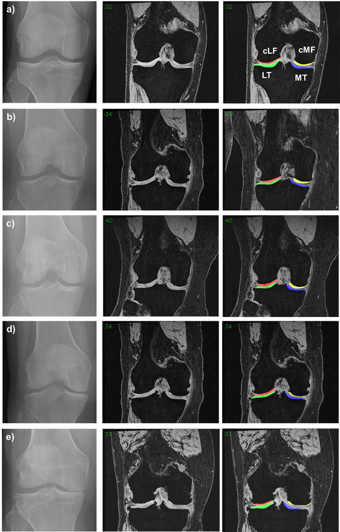
Online Figure 1. Knees from a OAI participants with different radiographic status: fixed flexion radiographs shown on the left, coronal MR images without segmentation shown in the middle; coronal MR images with segmentation of the femorotibial cartilages shown on the right.

a) Healthy reference cohort knee
b) Central KLG1 knee
c) Central KLG2 knee without JSN
d) Central KLG2 knee with JSN
e) Central KLG3 knee
MT = medial tibial cartilage; cMF = medial weight-bearing femoral cartilage; LT = lateral tibial cartilage, cLF = lateral weight-bearing femoral cartilage
