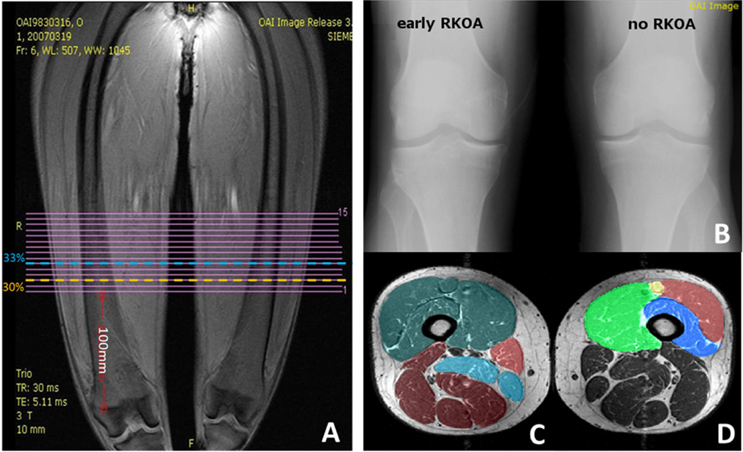
Figure 1.
A Coronal localizer image: 15 continuous axial slices of the thigh have been acquired. B. Participants (right top). For the current study we selected participants with early radiographic knee osteoarthritis (RKOA) in one knee and no RKOA in the contralateral knee on fixed-flexion X-rays (right top). C–D. Axial cross-sectional MRIs with segmented muscles. Anatomical cross-sectional areas of the quadriceps (pink), hamstrings (red), and adductors (yellow) have been segmented at 33% of femoral length (from distal to proximal) (C). ACSAs of the individual quadriceps heads vastus medialis (brown), vastus intermedius (turquoise), vastus lateralis (yellow), and rectus femoris (purple) have been segmented at 30% of femoral length (from proximal to distal) (D).

